Positive Health Online
Your Country

Osteopathic Approach to the Treatment of Facial-Maxillary Sinus Headache
listed in osteopathy, originally published in issue 206 - May 2013
Introduction
Anterior facial pain/headache caused by swollen sinuses usually results from infection both bacterial / viral or an allergic reaction, resulting in blockage to the drainage channels.
|
|
 |
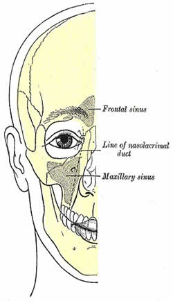
Drawing of Sinus Structures Anatomical Drawing of Facial structures
Signs and Symptoms
The patient usually experiences pain over the supra orbital ridge (frontal sinus), between the eyes (ethmoidal) which can be associated with anosmia and cheeks (maxillary). As the sinuses are innovated by the trigeminal nerve (CNV) pain can be felt in the mouth, upper teeth, hard palate, frontal, nasal and orbital areas. It is frequently reported as a dull facial ache to severe pressure and should not be confused with migraine headache.
Ultimately the sinuses drain via the sinus ostium an opening that connects a sinus to the nasal cavity. It is a tight area that tends to have a higher percentage of cilia than the surrounding mucosa.

Sinus Structure
If the sinus ostium is blocked, the entire sinus thus becomes a pathologic cavity or cyst like lesion. As mucus is accumulated and the sinus cavity has filled, the increase in intraantral pressure results in a thinning, displacement and, in some cases, destruction of the sinus walls. If the mucocele becomes infected, it is called a pyocele or a mucopyocele.
Factors which may predispose to developing sinusitis include: allergies, structural problems such as a deviated septum or small sinus ostia, smoking, nasal polyps, carrying the cystic fibrosis gene (research is still tentative); prior bouts of sinusitis as each instance may result in increased inflammation of the nasal or sinus mucosa and potentially further narrow the openings.
Differential Diagnosis
- Classical migraine;
- Trigeminal neuralgia;
- Temporomandibular dysfunction;
- Tooth infection;
- Skull Fracture;
- Facial maxillary carcinoma.
Evaluation and Diagnosis
Pain, swelling and tenderness is felt over the affected sinus. Nasal discharge may be yellow, green indicating infection or clear may indicate a basilar skull fracture so any history of head injury should be noted. By using a pen light the operator should check for reduced transillumination of the affected sinus.
Also assess movements of the TMJ and cervical spine for associated dysfunction, along with the anterior neck musculature; hypertonicity here could obstruct drainage from the head and neck causing back/flow pressure into the head.

Sutures of the skull
Techniques to Assist Sinus Drainage
Extra-oral Technique

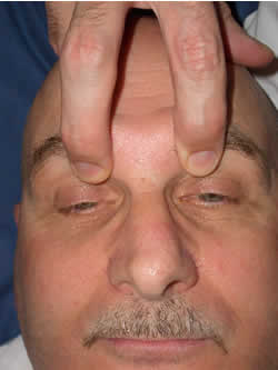
Frontal Lift to coronal suture
Procedure
- With the patient supine the operator stands at the head of the couch;
- With the head supported to avoid extension, the operator places the finger pads of his index and mid fingers under the patients supra orbital ridge, care being taken not to make contact with their eye
- Gentle pressure is then applied to lift the frontal bone from the nasal and ultimately the ethmoid, which can be re-enforced by the operator gently griping the upper aspect of the nose with their thumb and index finger. Thereby opening the sinus ostium;
- The technique can be re-enforced by holding back on the nasal bone;
- Re-assess.
Intra-oral Technique
 |
 |
Inhibition to the base of the maxillary sinus
Procedure:
- With the patient supine and head and neck supported to avoid extension;
- The patient is asked to open their mouth fully;
- Using a gloved hand the operator inserts his index into the patient’s mouth following the line of the hard palate until the junction is felt between the hard and soft palates;
- Using an upward pressure the operator gently presses up into the soft palate;
- This creates an increased pressure on the base of the facial-maxillary sinus directing the contents of the sinus up and out.
Comments:
-
No Article Comments available
